How the Hands-On Abdominal Ultrasound class typically unfolds.
Our abdominal course unfolds as a systematic, high-yield survey of visceral imaging, Doppler interpretation, and clinical reasoning.
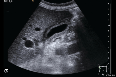
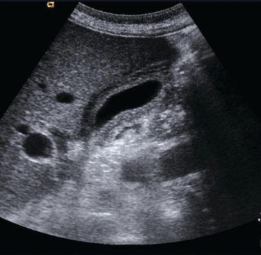
Early sessions focus on image ergonomics and machine optimization, giving learners tactile confidence and geometric orientation to three-dimensional anatomy across the abdomen. From there, we move through a comprehensive organ-by-organ protocol, teaching learners to interpret anatomy, echogenicity, Doppler blood flow, and physiologic relationships with clarity.
Hands-on practice is continuous. Learners repeatedly apply Doppler to vascular structures, correlate findings with pathology, and learn how to document each organ and vascular bed in a methodical way.
Concepts are revisited often so learners deeply connect pattern recognition to clinical decision-making. Optional independent Scan Lab time reinforces every technique, building lasting competence you can carry straight to the bedside. And after class adjourns, our lifetime mentoring stays at the bedside with you.
Training Clinicians Worldwide Since 1981.
Testimonials reflect individual learning experiences. Growth in skill and confidence develops through guided training, continued practice, and personal commitment.
Contact
972 | 353-3200 USA Central Time
Course Campus:
4300 Wingren Drive
Irving (Las Colinas), Texas 75039
Mail | FedEx Address:
Box 101
Colleyville, TX 76034
Courses & Learning
Ultrasound Physics
Cardiovascular Hemodynamics
EKG- From Atoms to Arrhythmias