Hands-On Transvaginal Pelvic Ultrasound Imaging & Doppler
A single-day guided experience that develops the skill to evaluate pelvic anatomy and physiology accurately, confidently, and with disciplined clinical reasoning.
“I was new at transvaginal ultrasound imaging and I feel comfortable doing the scan now.”
-Tina Cunningham
Get here. Learn It. Practice it. Do it. Serve.
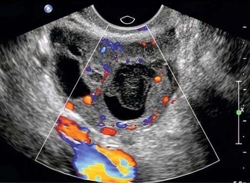
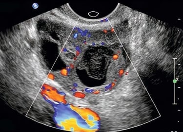
Imaging the Pelvis with Clarity.
This is where precision, confidence, and women's care converge. We’ve developed one of the most effective live, hands-on transvaginal ultrasound training experiences available today. In this course, you’ll learn how to perform a complete, efficient, and accurate pelvic exam—from the first image to the final Doppler sweep.
By connecting sonographic anatomy, physiology, and pathology, you’ll strengthen your understanding of both normal and abnormal findings—and build confidence in your patient care.
Personal mentoring support continues after class, always free of charge.
Why This Course Matters For You—Because Evasive Sonographic Anatomy Drives Clinical Decisions.
Pelvic imaging brings technical demands and clinical responsibility together. This hands-on course matters because it trains you to approach deep anatomy with precision, interpret subtle cues within context, and make confident calls that directly affect care planning.
We impart a method of precise anatomic dead reckoning to cut your time and seal your confidence. Through guided scanning and focused repetition, you’ll develop the skill and judgment to move beyond uncertainty to actionable insight.
What Makes This Course Different.
Taught as spatial navigation, not pattern memorization – Develop true 3D anatomic understanding from 2D images, eliminating probe-guessing and hesitation.
Hands-on dominance from the first moment – Guided, deliberate scanning forms the foundation of learning rather than extended lecture or observation.
Instruction adapts to the learner – Small class size allows faculty to adjust teaching in real time to your experience and pace.
Confidence that transfers immediately to practice – Leave with a disciplined, repeatable approach you can apply the very next time stakes are high.
How the Course Typically Unfolds.
The class is structured to build confidence, clarity, and clinical precision in every learner from the first scan.
We begin with respectful patient approach and machine optimization, ensuring that learners feel grounded in both technical and human aspects of care. A rapid, sequential pelvic protocol is introduced early, so each scan has purpose and flow.
Hands-on practice emphasizes complete, systematic exams of the uterus, ovaries, adnexa, and Doppler assessment where indicated — each with faculty coaching on image optimization and interpretation. Brief reflective discussions bridge anatomy, physiology, and normal vs expected pathological findings.
Scanning sessions are designed to build familiarity and muscle memory, progressing from basic views to more advanced pelvic documentation. Independent lab time reinforces skills at your pace. By the end of the day, you’ll walk away with a clear, efficient protocol and interpretive logic you can immediately apply in women’s health settings
Tuition—Your Learning Investment.
Smart Career Growth— Zero Debt→
This investment reflects the micro-class size, intensity of hands-on instruction, detailed course materials you'll reference forever, unlimited scan-lab access, and lifetime post-course mentoring. Hot breakfast and light lunch are included daily.
Tuition $1000 (USD) Monday- Thursday.
9am - 4pm, adjourn by 3pm on Thursday.
Scan Lab open 24 Hours during class.
If tuition is a concern, we’ve already thought about that.
Download this guided Employer-Sponsorship Agreement Kit→ that lets your workplace pay for the course while you agree to stay for a defined time afterward. It’s one of the easiest ways to advance your skills without taking on new expenses.
Most attendees say the Course pays for itself in just a few weeks of bedside application.
About CME Credit.
This course does not offer CME credit—by design.
That choice allows us to teach with complete flexibility, adapting instruction to your background, pace, and clinical goals rather than a committee's preset agenda.
Our focus is hands-on skill development, clinical reasoning, and lasting bedside confidence. Many clinicians choose to pair this experience with separate CME activities that align with their own professional requirements. If we can help direct you to such low- or no-cost CEM resources, we'lll be delighted to assist.
What Our Participants Said.
The overwhelming majority described the experience as excellent, transformational, and more effective for them than other national ultrasound courses.
Students repeatedly mention they learned more in a few days than in months elsewhere.
What Our Participants Said.
The overwhelming majority described the experience as excellent, transformational, and more effective for them than other national ultrasound courses.
Students repeatedly mention they learned more in a few days than in months elsewhere.


You've Got Questions?
Let's face them together.
Your decision is transformational. Ask us anything and get a straight answer. We want to address your every question, because you and what you’re about to do are important.
Your Next Step Is Closer—and Brighter Than You Think.
You've come so far already— don't quit.
You’ve considered the most demanding parts: the time apart from the people who matter, the disruptions of travel, the honest moments of doubt, and the uncertainty we all feel when something truly matters. Here, you are safe. Here, you are supported. And what you gain will elevate you, your family, and your patients for years to come.
It would be easier—and much more hollow—to simply turn on the TV or computer and watch a video. But every lasting accomplishment comes by doing. You don’t learn ultrasound skills sitting on your hands.
Start your success now—without hesitation:
Download your free Anatomy Charts and Reference Guides→
Connect directly with Keith→ on LinkedIn for personal guidance
Request to join our private Alumni Community→ on Facebook.
Your Journey is already underway.
We’ll meet you in the Scan Lab soon—and at the Bedside for the rest of your career....
“You related the information and practice directly to exam protocol.
Very effective.”
-Steven J. Pritchard
Training Clinicians Worldwide Since 1981.
Testimonials reflect individual learning experiences. Growth in skill and confidence develops through guided training, continued practice, and personal commitment.
Contact
972 | 353-3200 USA Central Time
Course Campus:
4300 Wingren Drive
Irving (Las Colinas), Texas 75039
Mail | FedEx Address:
Box 101
Colleyville, TX 76034
Courses & Learning
Hands-On Clinical Courses
Ultrasound Physics
Cardiovascular Hemodynamics
EKG- From Atoms to Arrhythmias
Privacy Policy | Terms of Use. | Disclaimer
We’re committed to accessibility. View our Accessibility Statement.
The most powerful technology in medicine
is the person using it.