Hands-On Vascular Ultrasound Imaging & Doppler
The four-day guided experience that trains you to read blood flow as a living language—
revealing disease long before and immediately after symptoms appear.
“They empowered me with knowledge that can never be taken away.”Sandra M. Alvarado
Get here. Learn It. Practice it. Do it. Serve.
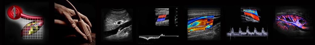
Reading blood in motion.
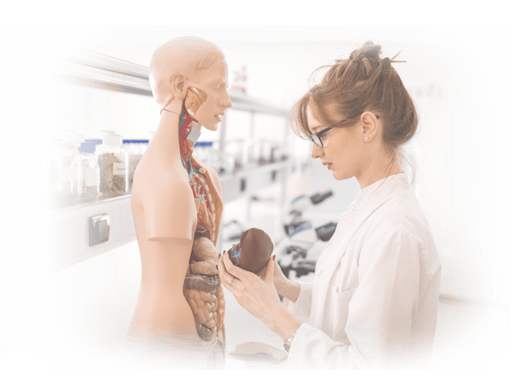
Build your vascular ultrasound career on the shoulders of the persons who pioneered it and helped frame the standards for it. Vascular ultrasound doesn’t have to feel complicated or overwhelming. In our hands-on, head-to-toe vascular program, you’ll discover just how clear, structured, and confidence-building this field can be when you learn it through a practical, step-by-step approach. From your very first hour, you'll be scanning with purpose—guided by a protocol that makes sense, supported by instructors who stand beside you both in class and long after you head home.
Across four immersive days, you’ll learn a unified, repeatable workflow that applies to carotid, abdominal, and peripheral arterial and venous ultrasound. You’ll see how all vascular exams share the same hemodynamic foundations, and you’ll learn to interpret every finding with both accuracy and clarity. Thousands of clinicians from every specialty have trusted this course to launch or advance their vascular competency—and you’ll soon understand why.
"I Don't Scan Vessels, I Scan People"—Why This Course Matters For You Now
Blood flow is physiology in motion, and static assumptions can mislead. This course matters because it teaches you to apply Doppler and grayscale imaging with intentional technique, interpret flow patterns with physiologic context, and recognize pathology as meaningful deviation rather than incidental noise.
The hands-on format gives you repeated live practice, immediate correction, and the competence to use vascular ultrasound as a dependable component of your clinical decision-making. And if your focus is on the certification exams, this is the definitely the place to prepare your mind.
What Makes This Course Different
Taught as a clinical skill, not a memorized protocol.
Learn why each image and Doppler assessment matters, so you can adapt confidently when anatomy, pathology, or patient condition doesn’t follow the textbook.
Physiology-driven, bedside-ready instruction.
Understand real blood-flow behavior and hemodynamics—not just probe positions—so findings make immediate clinical sense.
Immediate recognition of normal and abnormal flow.
Train your eye to identify waveform patterns quickly, troubleshoot poor signals, and reason through complex vascular findings in real time.
Hands-on scanning with direct faculty guidance.
Small classes keep instructors at the machine with you, offering continuous feedback as you scan on live models—not from across the room.
Repetition that builds instinctive confidence.
Scan repeatedly until technique, pattern recognition, and decision-making become second nature rather than something you have to think through.
Extended access to practice time.
Our scan lab remains open beyond formal class hours, allowing you to refine technique and solidify understanding at your own pace.
Confidence that carries into unfamiliar exams.
Leave prepared to approach new patients, new pathologies, and complex vascular questions with clarity and control.
How Each Professional Benefits By Attending
Sonographers and Echocardiographers
Vascular imaging elevates your entire scanning skill set. This course strengthens your ability to interpret hemodynamics, recognize abnormal flow patterns, and perform precise Doppler angle correction across arterial and venous systems. You’ll gain a clear, protocol-driven approach that complements your cardiac and general scanning experience—expanding your clinical versatility and making you an invaluable part of any diagnostic team.
Sonography Graduates
This training gives you exactly what employers look for: real scan time, vascular logic, and a repeatable protocol you can demonstrate with confidence. You’ll learn how to acquire, analyze, and document vascular studies correctly from day one. Your résumé becomes instantly stronger, and you’ll be ready the moment someone says, “Show me how you scan.”
Allied Health Professionals
Vascular imaging is one of the most widely needed—and misunderstood—skills across healthcare. This course gives you a step-by-step, clinically defensible workflow to evaluate perfusion, DVT risk, arterial stenosis, and more. Whether you’re expanding your clinical role or adding ultrasound to your existing duties, you’ll leave with a clear method you can apply immediately and safely.
Critical Care Providers
Rapid vascular assessment saves time, changes management, and improves patient outcomes. You’ll learn to perform focused bedside evaluations of carotid stenosis, major DVT, and perfusion-limiting arterial disease with confidence—making you faster, more accurate, and more decisive in the moments that matter most. This aligns seamlessly with point-of-care protocols and modern critical care workflows.
Physicians Preparing for the RPVI Exam
Nothing accelerates RPVI mastery faster than hands-on scanning combined with clear hemodynamic logic. This course gives you the experiential foundation that transforms theory into intuition—instantly improving your waveform interpretation and dramatically shortening your daily reporting time with clearer analytic criteria.
Anesthesiologists & PACU Teams
Understanding sonographic vascular images and flow patterns at the bedside enhances pre-op screening, post-op complication assessment, and targeted anesthesia planning. You’ll learn how to rapidly evaluate perfusion, venous patency, thrombotic risk, and postoperative vascular concerns, improving collaboration with surgical teams and elevating patient safety.
Medical Device & Research Professionals Research, Engineering, Marketing, Sales, Management, Applications, Service Engineers
Hands-on exposure to real vascular imaging challenges unlocks deep insight into how clinicians actually acquire, optimize, and interpret arterial and venous studies. You’ll leave with the practical understanding needed to better design, support, and communicate around vascular devices, software, and instrumentation—with far greater accuracy and credibility.
We Teach the Standards—Your Way
There’s a difference between what we teach and how we teach it. The content never changes—it’s grounded in decades of practice and the standards clinicians rely on. But the delivery must adapt in real time to ensure the content truly sticks. We continually read the room, adjusting moment by moment to each learner’s background, comfort level, and natural way of receiving complex ideas. Every essential concept is presented multiple times, in a variety of ways, so key principles take root quickly and stay with you. Here’s how a typical class may flow.
Day 1
Carotid & Vertebral Arteries
Begin with the complete carotid duplex protocol, learning exactly why each step matters. You’ll master image optimization, artifact recognition, and hemodynamic interpretation while exploring extracranial cerebrovascular disease with clarity and confidence.
Day 2
Lower Extremity Arterial System & TCD
Explore targeted MCA transcranial Doppler and apply carotid principles to the arteries of the legs. You’ll complete native and post-operative arterial protocols and pair duplex with physiologic testing to evaluate limb perfusion with clinical authority.
Day 3
Venous Imaging & Abdominal Vascular Protocols
You’ll perform complete deep and superficial venous exams for DVT and venous insufficiency. Then we expand into abdominal vascular ultrasound—including aorta, IVC, hepatic vasculature, and renal arteries—with diagnostic criteria made understandable and actionable.
Day 4
Upper Extremity Arterial & Venous Exams
Complete duplex protocols for both arterial and venous systems in the upper extremities. We end with focused practice time tailored to your individual interests and application goals.
Throughout the Week
Your on-site Scan Lab remains open around the clock for independent practice—on yourself or with classmates—so your confidence grows with each repetition. Every step, every protocol, and every concept is explained in full detail in your 200-page course notebook, a resource you will return to for the rest of your career. And when you return home, our personal support continues—free and in perpetuity.
What You'll Learn Clinically
We shrink the class size on purpose, ensuring we can walk through every critical topic together while still giving you the freedom to ask, explore, and discuss whatever you need.
How to perform every major vascular protocol—with a unified, repeatable approach
A simple three-dimensional strategy to obtain high-quality vascular images quickly and consistently
How to evaluate aneurysm, dissection, flow-limiting stenosis, and advanced hemodynamic changes in native vessels, grafts, stents, and dialysis fistulae with clarity
Renal artery and abdominal vascular assessment without diagnostic compromise
Deep and superficial venous mapping for DVT and venous valvular insufficiency
Understanding waveform changes throughout the arterial cycle and what they mean clinically
Raynaud’s assessment, thoracic outlet interpretation, and cold sensitivity testing
How to think through any ultrasound registry exam question using reasoning—not memorization.
Our Approach.
Ultrasound skill is built through guided experience, not passive observation. Instruction centers on live scanning, immediate feedback, and repeated practice—so technique, interpretation, and clinical reasoning develop together.
We teach in tiny groups to allow faculty to adjust instruction in real time, responding to each learner’s background, pace, and questions. Concepts are revisited from multiple angles, with real-world explanations, helping understanding take root quickly and remain reliable under clinical pressure.
The emphasis is not on memorizing images or following rigid protocols, but on learning how to think at the bedside—recognizing patterns, understanding physiology, and making deliberate decisions that translate directly to patient care.
There's no written test—just continuous encouragement and hands-on coaching to help you grow.
Tuition— Your Career Investment
Smart Career Growth— Zero Debt.
This investment reflects the micro-class size, intensity of hands-on instruction, detailed course materials you'll reference forever, unlimited scan-lab access, and lifetime post-course mentoring. Hot breakfast and light lunch are included daily.
Tuition $2900 (USD) Four Days
9AM - 4PM, Adjourn by 3PM final day
Scan Lab open 24 hours during the Course
If the cost is a challenge, we’ve already thought about that.
Download this Employer-Sponsorship Agreement Kit→ that lets your workplace pay for the course while you agree to stay for a defined time afterward. It’s one of the easiest ways to advance your skills without taking on new expenses.
Tuition covers everything you need to learn comfortably and confidently—your formal discussions and hands-on practice, detailed course materials you’ll keep forever, a hot breakfast and light lunch onsite, and continued support after the course so you never feel on your own.
Most attendees say the Course pays for itself in just a few weeks of clinical application.
What Our Participants Said.
The overwhelming majority described the experience as excellent, transformational, and more effective for them than other national ultrasound courses.
Students repeatedly mention they learned more in a few days than in months elsewhere.
About CME Credit
This course does not offer CME credit—by design.
That choice allows us to teach with complete flexibility, adapting instruction to your background, pace, and clinical goals rather than a committee's preset agenda.
Our focus is hands-on skill development, clinical reasoning, and lasting bedside confidence. Many clinicians choose to pair this experience with separate CME activities that align with their own professional requirements. If we can help direct you to such low- or no-cost CEM resources, we'lll be delighted to assist.
You've Got Questions?
Let's face them together
Your decision is transformational. Ask us anything and get a straight answer. We want to address your every question, because You and what You’re about to do are important.
Dallas, Texas, USA
Central Time UTC: -06:00 (CST)
Your Next Step Is Closer—and Brighter—Than You Think
You've come so far already— don't quit.
You’ve considered the most demanding parts: the time apart from the people who matter, the disruptions of travel, the honest moments of doubt, and the uncertainty we all feel when something truly matters. Here, you are safe. Here, you are supported. And what you gain will elevate you, your family, and your patients for years to come.
It would be easier—and much more hollow—to simply turn on the TV or computer and watch a video. But every lasting accomplishment comes by doing. You don’t learn ultrasound skills sitting on your hands.
Start your success now—without hesitation:
Download your free Anatomy Charts and Reference Guides→
Connect directly with Keith→ on LinkedIn for personal guidance
Request to join our private Alumni Community→ on Facebook.
Your Journey is already underway.
We’ll meet you in the Scan Lab soon—and at the Bedside for the rest of your career....
Training Clinicians Worldwide Since 1981.
Testimonials reflect individual learning experiences. Growth in skill and confidence develops through guided training, continued practice, and personal commitment.
Contact Us
972 | 353-3200 USA Central Time
Course Campus:
4300 Wingren Drive
Irving (Las Colinas), Texas 75039
Mail | FedEx Address:
Box 101
Colleyville, TX 76034
Legal & Administrative
Privacy Policy
Accessibility Statement
© Keith Mauney & Associates Ultrasound Training Institutes MMXXVI. All rights reserved.
Courses & Learning
Upcoming Course Dates
Your Skills-Building Experience
Hands-On POCUS- Point of Care Ultrasound
Hands-On Adult Echocardiography
Hands-On Transvaginal Pelvic Ultrasound
Ultrasound Physics
Cardiovascular Hemodynamics
EKG- From Atoms to Arrhythmias