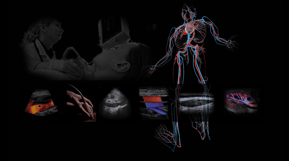
Precise, protocol-driven hands-on tutorial in total-body vascular ultrasound….
You’re about to learn that Vascular ultrasound training doesn’t have to be complex or intimidating: It’s simply the application of several basic principles to a lot of different protocols. You’ll be led in this Class along a path that will help you competently perform- and intuitively understand- all the findings and principles underlying each one. We’re uniquely qualified to escort you to your launch point quickly, and we’ll remain in your support whenever you need, long after you leave.
Everyone wonders what can be done in just four days here. We’d recommend you ask around the many thousands of persons who’ve passed our way since the beginnings of vascular ultrasound.
In the first hour, we’ll begin the complete carotid duplex protocol and explain in the clearest of terms the why behind each step. From there we’ll move down the body and analyze every accessible artery and vein while setting forth the unbreakable laws of blood flow.
Every word and process is directed to your successful passing of your credentialing exam and- more importantly- your bedside patient exams for life.

Day 1 You learn the extracranial carotid and vertebral duplex exam protocol and how to expand upon it when necessary. You’ll master imaging controls to minimize artifacts and learn how to spot and eliminate them. We’ll apply the principles of hemodynamics at every stage of vessel disease to the use and limits of color, spectral, and power Doppler. And in the process, we’ll marry it all with the spectrum of cerebrovascular disease.
- Evening You’ll have optional reading in your course materials and access to the on-site scan lab for practice on yourself and/or class peers.
Day 2 You’ll see the simple technique for targeted MCA assessment in transcranial Doppler. We’ll apply the knowledge mastered in the carotids to the arteries of the legs and execute the protocol for native and postoperative cases. We’ll discuss the combined roles of the duplex with physiologic tests to fully evaluate total limb perfusion. And we’ll set in motion the complete clinical approach to LE arterial disease so you can think and work with independent authority.
- Evening The on-site scan lab is open for your independent practice and optional reading will set the framework for deeply applied arterial hemodynamics.
Day 3 We’ll complete the deep and superficial venous protocols for thrombosis and valvular incompetence in the legs. We’ll introduce the aortic, vena cava, hepatic and detailed renal vascular protocols and the diagnostic criteria in each.
- Evening Optional assigned practice on calf vessel imaging in the scan lab. Continued independent practice.
Day 4 Full upper extremity arterial and venous assessment: duplex protocols and discussion of integrated physiologic testing. Further focus and guided practice on your topics of interest.
Who Will Benefit
Allied Health Care Providers
This vascular ultrasound training markedly advances professionals from all specialties to broaden and deepen their talents and open doors to lifelong opportunities. These include nurses, radiology technologists, and physicians of all specialties who will be scanning and/or solely interpreting vascular ultrasound. Foreign national physicians who find it impractical to re-board in the USA or Canada have found this a valuable path to expressing their expertise.
This Course will not confer the requisite 12 months of clinical experience required to apply for credentialing, but it will give you a particular edge at the start, when your potential Director says, “Now, let me see you scan….”
[Credentialing]
ER & Critical Care Providers: POC-Vascular Ultrasound
This ultrasound training is vastly deeper than the training your colleagues got in Residency. You’ll be able to evaluate immediately carotid patency and critical stenosis in the Stroke Response Team setting. Likewise, large vessel DVT and ambiguous claudication. The Point of Care Ultrasound Credentialing Academy has the tests to certify your skills (and generate revenue), elevating your own service to save time, costs, and improve outcomes.
Anesthesiologists & PACU Team: Targeted Transcranial and Carotid Assessment
Pre-op assessment of these vessels and intra-/post-operative recovery can provide instant answers that will create memories that will last a lifetime. If you wish to focus exclusively on these applications, inquire as to whether and how to attend only the first two days. These skills can also be taught to your entire team at once at your own site on any schedule that works with your flow. Let’s discuss your goals.
Sonography Students & Graduates:
Expand & Expound On Your Ultrasound Training
Many of the Students and Graduates of the hundreds of General Ultrasound programs will find far more job opportunities with vascular ultrasound training on your resume. Many students in this Class undertake it to build their foundation and/or expand upon it. Regardless of prior experience, we’ll be able to take you farther and deeper to prepare you for your future practice and credentialing. Your investment will pay out substantially when your potential Director says, “Let me see you scan.“
Research & Medical Ultrasound Device Professionals
No one in the field of medicine today has the depth and breadth of experience with ultrasound training for the Medical Device Industry as us. Over many years we’ve worked closely with nearly all ultrasound device manufacturers to steer and refine their products for clinical focus. We’ve also consulted with some of the largest Research Center on earth to help structure their work. As a Research Scientist, you’ll be able to identify and measure virtually any element of cardiovascular function. If you’re a Medical Device Professional- whether in-house or in the field, you’ll be better able to build, market, and sell your instrument with a unique competitive edge in the Service of many.
Are You New to Health Care?
Our ultrasound training focus is to take the practicing clinician and escort her or him to vastly greater hands-on ultrasound protocol and analysis skills in record time.
If you’re entering healthcare for the first time, you should consider your long-term goals and opportunities.
In North America, you can apply to any of hundreds of accredited schools (18+ months duration) and upon graduation immediately undertake your formal credentialing exam.
You’ll find the US Government’s most authoritative and current overview of the ultrasound career field here.
Topics
The class is strictly small so we can spend time on the topics we need to cover and all the ones you want to discuss:
- Putting together the complete vascular protocols: from the heart to the capillaries and back
- The three-dimensional secret to quickly obtaining the best possible vascular image
- The one simple technique (no one uses) to take 3 years off your mastery curve
- Abdominal and pelvic vascular ultrasound, from the aortic arch to the pelvis with focus on aneurysm, dissection, and all upper abdominal arterial and venous implications
- Renal assessment expedited without diagnostic compromise
- The single most powerful (and often overlooked) factor that can shut down the kidney in as little as twenty minutes
- The complementary role of physiologic testing and its unique contribution in end-stage arterial vascular disease, necessitating urgent intervention
- DVT: the single criterion for ultrasound diagnosis
- Controversies surrounding the limited LE DVT exam
- Carotid IMT: where we are in early patient screening management
- The complete 4-vessel protocol for carotid and vertebral artery assessment
- How to deal with the diagnosis and urgent management of subtotal vs. total carotid artery occlusion
- Neurology for the cerebrovascular ultrasound technologist: a brief and memorable review
- Understanding the statistical measures of diagnostic test accuracy for QA management
- How to analyze and document every pathologic finding: taking the right steps, using the right words.
- How to think your way through any Registry Exam question you might ever face.
- Next steps: How to reenter your workplace and maximize your next six months in the Field.
Objectives
Our approach is totally focused on the patient diagnosis. We are deeply familiar with virtually every ultrasound machine and the manufacturer’s rationale behind its design, features, and functions. No faculty members have any commercial interests or participation that might influence course content.
There is no formal test in this class: we evaluate you continuously and offer positive feedback and gentle corrections throughout. Upon completion of this activity, and through continued review, you should be able to:
- Quickly conduct a thorough, targeted patient history and physical/neurological inspection to obtain pertinent contextual information to amplify any test findings.
- Perform a complete duplex ultrasound exam of the cervical carotid and vertebral artery system; record and present all measurements of image and Doppler data.
- Identify, avoid, and correct image and Doppler artifacts through the use of machine controls and operator techniques.
- Chart the vascular anatomy and physiology of blood flow from the aorta to the brain and Circle of Willis.
- Describe the pathogenesis of atherosclerosis and relate it to the ultrasound-rendered findings of plaque morphology.
- Discuss the interrelationship of living plaque morphology and diameter reduction in the potential genesis of TIA and/or stroke.
- Set up monitor controls on any machine to maximize the display of the system’s input dynamic range, optimized for the human eye.
- Locate, explain, and make full use of post-processing controls to amplify soft tissue definition and relate the findings to histologic composition.
- Discuss the pertinent rules of hemodynamics and cardiac function applicable to the carotid and vertebral artery exam.
- Discuss the influence and clinical significance of each of the following on the accuracy of the duplex vascular exam:
- Doppler angle of incidence
- Angle correction factor
- Beam incidence and input dynamic range for both imaging and Doppler
- Biplane vs. triplanar imaging
- Doppler sample gate size
- Color vs. spectral Doppler sensitivity to slow flow and poor S/N ratios
- subtotal vs. total vessel occlusion
- clot echogenicity over time
- collateral flow.
- Demonstrate proficiency in auscultating cervical and orbital bruits and differentiate from a transmitted cardiac source.
- Perform Doppler assessment of any vessel using proper angle correction and display controls to maintain compliance with ICAVL Standards.
- Communicate all findings in a concise summary using standardized terminology and identify scenarios that require immediate physician attention.
- Acquire and identify Doppler signals from the proximal, mid, and distal MCA segments and apply pulsatility and resistivity indices to them
- Chart the vascular anatomy and discuss the physiology of blood flow to the legs and arms from the aorta to branching smaller vessels en route to the toes and fingers, through the capillaries, and back through the deep and superficial venous systems
- Describe the effects of arterial narrowing on each component of the normal Doppler spectral waveform in the peripheral arterial cardiac cycle
- Perform a complete ultrasound exam of the lower and upper extremity arteries, recording and presenting all measurements of image and Doppler data
- Perform and document a complete survey of the lower and upper extremity deep venous system in cases of suspected DVT and or venous valvular incompetence
- Map the greater and lesser saphenous veins of the leg, noting all principal branches communicating with the deep venous system
- Document the location(s) and classify the severity of deep and/or superficial venous valvular incompetence from thigh to ankle
- Map the arterial and deep/shallow venous vasculature of the arms from the aortic arch to the fingertips and back.
- Discuss the basis and protocol for assessment of nerve vs. vascular thoracic outlet syndrome.
- Discuss the pathophysiologic spectrum underlying Raynaud’s syndrome, the protocol for cold sensitivity testing, and the clinical ramifications of test findings.
- Demonstrate the protocol for the Allen test and explain the basis for normal and altered findings.
- Discuss the process and protocol for pre-and post-dialysis graft placement and/or revision
- Access current information regarding practice standards.
Tuition
$2600. Four days, 9 am-4 pm. Adjourns by 3 pm on the final day. On-site Scan Lab open 24 hours for independent practice. Your tuition includes your complete learning experience, printed course materials, and post-conference support in perpetuity. Breakfast and light lunch is provided daily.
CME
Your class experience is predominantly hands-on and the content is tailored hour-by-hour to both your specialty and experience. In this live interactive process, nothing is formulaic and fixed, as specified by the many varied CME accreditation bodies. Thus, we do not award formal Credit Hours, though your individualized experience here will advance your clinical skills dramatically. Presently, ultrasound credentialing prerequisites require Clinical Experience Hours before application for an exam. Ongoing CME is now specific to your registry specialty after you’re credentialed. To this end, we’ll always direct you to the most appropriate free and low-cost traditional CME credit activities available online.