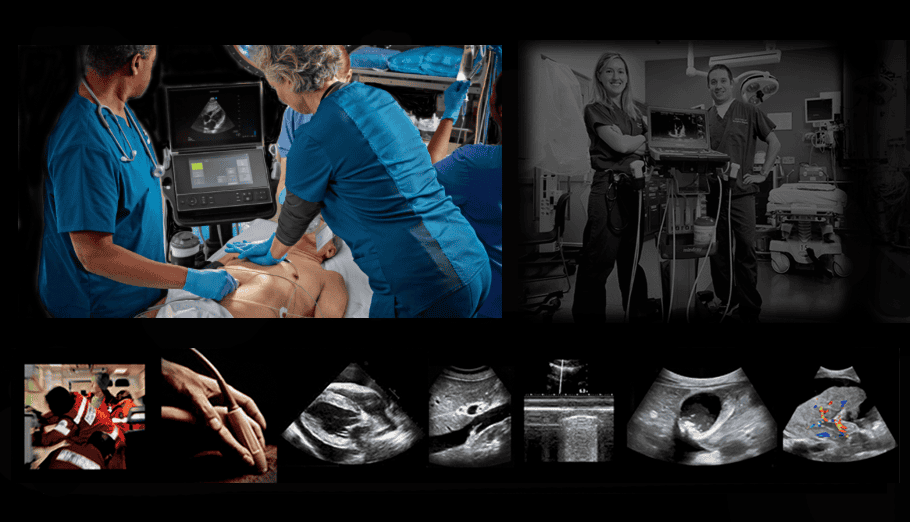
Put Real-Time Emergency Ultrasound Information in Your Own Hands
Gain authority with the complex POCUS instrument as you apply it to the complete protocols for emergency and critical care ultrasound. Learn and practice the best technique for ultrasound-guided vascular access. Hands-on-intensive ultrasound tutorials with lectures and one-on-one discussion to build your complete protocol to acquire and analyze all images. For all specialties and experience levels. 24-hour Scan Lab.
The most in-depth introduction to bedside ultrasound possible. Requested by the Attending Faculty at Harvard Medical School and the Mayo Clinic, this course goes deeper than just a protocol all in a weekend. Clear your understanding of ultrasound-guided vascular access. It all serves as the platform for your further competence in any clinical application. This is a very pleasant and practical, low-pressure approach to the complex science of acoustic and electronic engineering physics principles underlying bedside ultrasound. It cuts through the clutter and theory to make clear sense of all controls, principles, and findings.
Our focus is on the protocols and proper system controls for all current bedside applications. We start at the bedside in the first hour and continue throughout the course. After hours the Scan Lab is open for your optional self-guided practice of technique and machine controls on yourself or your peers from class. You can even bring your own equipment to fine-tune your own use of it.emergency/

Who Will Benefit
ER & Critical Care Physicians, Nurses, and Paramedics
You’ll be able to evaluate immediately right and left heart function and make critical decisions long before the formal echo report arrives. CVP, tamponade and pulmonary hypertension can be evaluated confidently and fast as soon as you return from class.
Allied Health Professionals
There are no prerequisites for this class. If you’ll be returning to an active clinical site you’ll arrive back with a giant leap forward. This weekend will prove that you can do it, give you the protocols to begin and lay the foundation on which you can build a lifelong career. You’ll be able to evaluate immediately right and left heart function and make critical decisions long before the formal echo report arrives. CVP, tamponade and pulmonary hypertension can be evaluated confidently and fast as soon as you return from class.
Anesthesiologists & PACU Nurses
This course is the answer to confidently staffing the PACU. Both you and the nursing staff will function at your highest level after this class.
Veterinary Medicine
Canine ultrasound runs on the same track as humans and the low cost/ high-performance equipment now in the market allows you to leverage it to enhance your practice. You’ll gain confidence quickly and our post-class support will ensure it lasts.
Research & Medical Device Professionals
As a research scientist, you’ll be able to identify and measure precisely virtually any element of cardiac function. If you’re a medical device professional you’ll be able to better build, market and support your product with an extra competitive edge.
Topics
The class is strictly small so we can spend time on the topics we need to cover and all the ones you want to discuss.
- Ultrasound physics in a single sentence: how to organize–and remember–everything about it.
- The one simple thing that will take three years off your learning curve and add a decade to your career.
- The secret to quickly getting every standard echo view without having to think.
- Speed reading cardiac wall motion abnormalities: taking a year off the learning curve.
- How to ensure you’ve found (or know that no one can find) the abdominal aorta and IVC.
- The no stone unturned approach to AAA.
- Why IVC respiratory variation doesn’t count: the better way.
- The fastest approach to the FAST exam.
- The triple play approach for free fluid: how to “run the bases”.
- Proving and localizing pleural effusion and documenting pneumothorax & consolidation.
- One stick, period: how to target the subclavian and IJV in 3-D space with ultrasound guidance.
- Clot or not: Why ultrasound prediction of echogenic aging of thrombus is a myth.
- Make the machine tell the truth: how to absolutely avoid image artifacts.
- Make the machine work for you (even with automated user controls): three things every clinician must know every time.
- Tough-Guy Techniques to overcome body habitus and air: the secrets you were never told.
- How to analyze and document every pathologic finding: taking the right steps, using the right words.
- Next steps: How to maximize your first six months in this new field.
Objectives
Our approach is totally focused on the patient diagnosis. We are deeply familiar with virtually every ultrasound machine and the manufacturer’s rationale behind its design, features, and functions. Even so, no faculty members have any commercial interests or participation that might influence course content. There is no formal exam in this class. Learners are evaluated continuously and positive feedback is offered throughout. Upon completion of this activity, you should be able to:
- Describe the systematic process by which ultrasound is generated, detected and processed into an image for analysis.
- Relate the physical phases of the acoustic wave to tissue response with emphasis on the mechanical and thermal indices.
- Define and describe in layman’s terms each of the principal vocabulary terms pertaining to ultrasound physics and instrumentation.
- Demonstrate proper probe grasp and manipulation technique for proper ergonomics and maximum dexterity.
- Apply a tactile approach to scanning to maximize stereotactic feedback.
- Follow a systematic protocol for assessment of each of the following:
-
- perirenal and perispenic fluid
- pericardial effusion vs. hemopericardium
- pleural effusion
- pneumothorax
- global and regional myocardial function
- aortic and mitral valvular regurgitation
- normal vs. increased CVP
- hepatic and portal venous hypertension
- biliary obstruction and renal lithiasis
- acute extracranial carotid occlusion
- DVT
- vascular access.
- Translate 3-D anatomical relationships to 2-D reasoning and vice-versa by applying the core principles of abstract spatial reasoning.
- Incorporate a full-field method of critical conspicuity to evaluate the entire content of any image immediately.
- Describe the principal controls of the ultrasound imaging system and effectively use each to optimize findings.
- Demonstrate proper use of overall and selective gain, depth and focus to optimize the acquired image.
- Set up image display controls on any monitor to maximize input dynamic range to output display capability properly optimized to the human eye.
- Make full use of all chroma and achromatic post-processing controls to amplify soft tissue definition and relate subsequent findings to soft tissue composition.
- Perform Doppler assessment of any vessel or valve using proper angle correction and display controls to maintain compliance with ICAVL Standards.
- Perform a visual inspection of the machine to identify any potential electrical or biohazard.
Tuition
$900. Two Days (Saturday-Sunday), 9am-4pm, Adjourn3 pm Sunday. Scan Lab also open 24 hours for your independent practice. Your tuition includes your complete learning experience, printed course materials and post-conference support in perpetuity. Breakfast and light lunch provided.
CME
We designate this course a non-CME credit activity, meaning that the curriculum is not defined by the constraints of a fixed format. This is to your strategic advantage. Though the content meets and exceeds every academic guideline, we have over many years determined that flexibility in tailoring specific elements—and the delivery to each individual learner’s need and style—supersedes the restraints of a traditionally rigid, time-limited format. It also permits us to openly, honestly and independently without bias discuss virtually every manufacturer’s system features. Too, the broad ranges of clinical professional specialist organizations have developed multiple exclusive brands of applicable CME. We have chosen instead to focus on your lifelong bedside competence and strategic advantage. This decision has permitted us to make our classes microscopically small at the least tuition cost possible. We will be delighted to direct you to the most appropriate free and low-cost traditional CME credit activities available online.