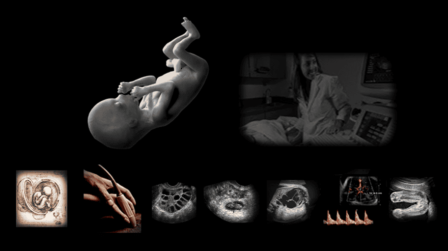
You’ll find your way through the fetus with competence, confidence…and respect….
Among the most rewarding, but challenging expressions of Women’s Care is obstetric sonography. This hands-on ultrasound training experience is totally focused on your tactile skills, abstract spatial reasoning, and pattern detection. Far more than learning to track a moving target, we’ll repeatedly practice the systematic protocol to make certain all accessible fetal structures are inspected, documented, and clearly communicated. Our approach, forged over forty years, will set you on the path to competence immediately and direct you toward- or return to the joy of making a difference.
Four days together at our bedside will change your trajectory forever. In the first hour, we’ll set forth the machine safety and optimization controls for both ultrasound imaging & Doppler. Then, our first pregnant subjects arrive and we work our way from the cervix to the fundus. We survey to first spot any urgent concerns, then methodically document fetal anatomy from head to toe. Between cases, we’ll formally present and discuss every element of the standard anatomic survey protocol, then return again and again to practice it.
When and where Doppler might have a role, we’ll safely and judiciously apply it and learn its powerful subjective and objective contribution. Woven into this continuous practice is our steady reinforcement of how to know, not just what to do and remember.

Day 1 We set forth the proper tactile approach to probe grasp to maximize dexterity and preserve ergonomic practice. We’ll demonstrate and discuss the three prime instrument controls that most powerfully affect the accuracy of your image. You’ll begin to apply our cervix-to-fundus protocol on theoretically normal pregnant subjects, varying from 12-36 weeks. Between cases we’ll segment the formal presentation of the precise fetal anatomy scan, beginning with the head.
Evening Your optional assignments include surveying your course materials to inventory your didactic resources. The on-site scan lab is accessible for your independent practice on yourself or in-class peers to practice fundamental techniques and machine controls.
Day 2 We begin with a discussion of your overnight questions and proceed further into the formal details of the fetal anatomy scan protocol emphasizing the fetal heart. At the bedside with our pregnant subjects, we’ll introduce the use of color, power, and spectral Doppler: how to optimize each and the valuable data that can be gleaned.
- Evening The on-site scan lab remains open for your optional continued practice on yourself or in-class peers.
Day 3 After discussing your overnight questions, we’ll discuss techniques to identify the RVOT & LVOT and how to inspect the IAS and IVS. In between scanning our pregnant guests, we’ll complete the analysis of the anatomy scan protocol and detail the survey of the adnexa for ectopic and heterotopic pregnancy. We’ll use color Doppler to evaluate placental integrity and impart the basis of the umbilical cord and fetal MCA spectral Doppler for evaluation of IUGR. By the end of the day, we’ll discuss how to survey the maternal kidneys and document their changes during pregnancy.
- Evening Assigned optional practice in the on-site scan lab: image and Doppler assessment of the kidneys, on yourself or in-class peers.
Day 4 Our morning begins with a formal presentation of the Biophysical Profile protocols, assessment of IUCD’s, and the transabdominal ultrasound contribution to Gyn evaluation in pelvic pain and bleeding. Our final scanning sessions allow your self-assessment and concretion of all learning.
As you prepare to return to your practice, we’ll prescribe the resources and connections vital to your continued development over the next six months. And when you leave, we’ll stand beside you with free support in perpetuity.
Who Will Benefit
Allied Health Care Providers
This hands-on OB ultrasound training gives professionals from all specialties the skills to broaden and deepen their talents and open doors to lifelong opportunities. These include Radiography and Respiratory Therapy Technologists, Traditional and Advanced-Practice Nurses, Midwives, and PAs. Foreign Physicians who find it impractical to re-board in the USA or Canada have found this a valuable path to expressing their clinical service expertise.
This Course will not confer the requisite 12 months of clinical experience required to apply for credentialing, but it will give you a particular edge at the start, when your potential future Director says, “Now let me see you scan….”
[Sonographer Credentialing]
Residents and Physicians
The hands-on ultrasound training you got in Residency is valuable; four days with us will build on top of it a skyscraper of future equity. You’ll save time and reduce risk as you oversee and sign off on your Sonographers’ work. And when they’re not in agreement with your clinical assessment, you’ll be able to quickly step in and resolve any discrepancy. Pocket ultrasound imaging with color and now spectral Doppler is advancing ultrasound as fast as its price is dropping. High-performance technology is now within reach of every individual clinician who elects to make it your own. This Class is the shortest and most effective path to your goal and our post-Class support ensures your future confidence and competence.
Sonography Students & Graduates:
Expand & Expound Your Ultrasound Training
Students and Graduates of formal General Ultrasound programs will find far more job opportunities with echocardiography training on your resume. Many of our colleagues in this Class undertake it to build their foundation and/or expand upon it. Regardless of prior experience, we’ll be able to take you farther and deeper to prepare you for your future practice and credentialing. Your investment will pay out substantially when your potential Director says, “Let me see you scan.“
Research & Medical Ultrasound Device Professionals
No one in the field of medicine today has the depth and breadth of experience with ultrasound training for the Medical Device Industry as us. Over many years we’ve worked closely with nearly all ultrasound device manufacturers to steer and refine their products for clinical focus. We’ve also consulted with some of the largest Research Center on earth to help structure their work. As a Research Scientist, you’ll be able to identify and measure virtually any element of cardiovascular function. If you’re a Medical Device Professional- whether in-house or in the field, you’ll be better able to build, market, and sell your instrument with a unique competitive edge in the Service of many.
Are You New to Health Care?
Our ultrasound training focus is to take the practicing clinician and escort her or him to vastly greater hands-on ultrasound protocol and analysis skills in record time.
If you’re entering healthcare for the first time, you should consider your long-term goals and opportunities.
In North America, you can apply to any of hundreds of accredited schools (18+ months duration) and upon graduation immediately undertake your formal credentialing exam.
You’ll find the US Government’s most authoritative and current overview of the ultrasound career field here.
Topics
The class is strictly small so we can spend time on the topics we need to cover and all the ones you want to discuss:
- Quick and forever orientation to any pelvic image: gravid or not
- The secret tactile tool that will take 3 years off your mastery curve and forever relieve your wrist strain
- The two-step process to launch and complete the head-to-toe fetal anatomy scan.
- The secret to optimize every image and Doppler finding without having to think
- System (and monitor) setting to standardize every image, every time.
- Landmarks, scan planes and trajectories for the fetal head: brain, posterior fossa& lateral ventricles, spine, chest & heart, stomach, kidneys & bladder, cord insertion, extremities, and gender
- The systematic approach to identifying the right and left heart outflow tracts: secrets of the echocardiographers
- Locating, grading and mapping the placenta and its uterine integrity
- Fine-tuning the measurement of AFI
- Cord and middle cerebral artery Doppler: analysis of resistance, pulsatility and systolic/diastolic indices in evaluation for IUGR
- Why the the maternal heart and kidneys matter and what to look for
- Systematic assessment of the adnexa in suspected ectopic or heterotopic pregnancy
- The non-gravid pelvis: Techniques to overcome body habitus and air- the secrets you might not have even thought of.
- How to analyze and document every pathologic finding: taking the right steps, using the right words.
- How to think your way through any Registry Exam question you might ever face.
- Next steps: How to reenter your workplace and maximize your next six months in the Field.
-
Objectives
Our approach is totally focused on the patient diagnosis. We are deeply familiar with virtually every ultrasound machine and the manufacturer’s rationale behind its design, features and functions. No faculty members have any commercial interests or participation that might influence course content.
There is no formal test in this class: we evaluate you continuously and offer positive feedback and gentle corrections throughout. Upon completion of this activity, and through continued review, you should be able to:
- Demonstrate a prioritized survey of hte gravid pelvis to observe any gross or suspected abnormalities which migh terminate further routine study and require physician attention
- Conduct a systematic ultrasound fetal anatomy scan and measure/document all pertinent anatomic structures.
- Locate and document the situs of the fetal heart with a focus on 4-chamber development, inflow and outflow tracts, septae, and cardiovascular structural integrity
- Estimate fetal gestational age using cranial, abdominal and extremity metrics
- Identify and correct common sources of operator error in acquiring the most accurate images for measurement
- Apply spectral Doppler measurements of cord and middle cerebral artery resistance index to reconcile IUGR in fetal development tracking small for dates
- Optimize and apply judicious use of color and/or power Doppler to evaluate for placental abruption
Tuition
$2600. Four Days, 9am-4pm; adjourn by 3pm on final day. The on-site scan lab is accessible during evening hours for your independent optional assignments Your tuition includes your complete learning experience, printed course materials and post-Class support in perpetuity. Breakfast and light lunch is provided.
CME
Your class experience is predominantly hands-on and content is tailored to both your specialty and experience, hour-to-hour. In this live interactive process, nothing is formulaic and fixed, as specified by the many varied CME accreditation bodies. Thus, we do not award formal Credit Hours, though your individualized experience here will advance your clinical skills dramatically. Presently, ultrasound credenaitling prerequisites require Clinical Experience Hours prior to application for exam. Ongoing CME is now specific to your registry specialty after you’re credentialed. To this end, we’ll always direct you to the most appropriate free and low-cost traditional CME credit activities available online.