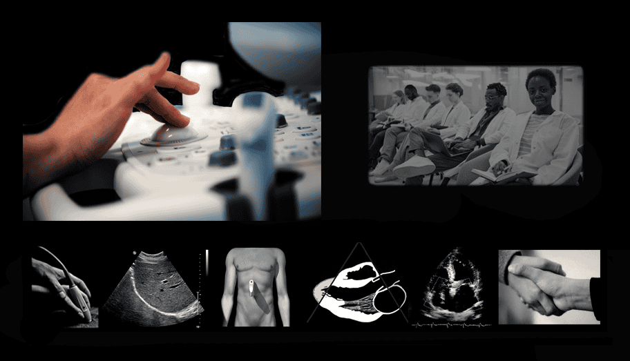
You can learn from a colleague for months and still not know the why behind what you’re doing. Or you can spend a powerful weekend with us and understand it immediately. You’ll stand on top of this granite foundation forever, for life. The technology, the economy, and the community are all moving into Point of Care (POC) Ultrasound and you need to gain this advantage now.
Just a weekend together will flex your trajectory forever.
In the first hour, we’ll set forth the real purpose, parameters, and limitations of ultrasound imaging in the body. Immediately, at the bedside, we’ll lead you through the tactile techniques, abstract spatial orientation, and conspicuity skills that’ll take years off your learning curve. You’ll decipher the vocabulary and know how to competently set out on your path to lifelong practice, confidence, and respect.
Class is micro-sized so we can spend time to meet you wherever you are and escort you to your Next Level. The on-site Scan Lab is accessible Saturday evening for your independent practice on yourself and/or class peers. And you’ll have a complete set of course materials that you can study to master the physics and instrumentation controls behind every ultrasound imaging and Doppler exam for life.
Stack Your Clinical Skills:
Your Giant Career Step Forward Starts Here

Class is micro-sized so we can spend time to meet you wherever you are and escort you to your Next Level. Our on-site Scan lab is accessible Saturday evening for you inde[endent practice on yourself and/or class peers. And you’ll have a complete set of your own course materials that demystify all the physics and instrumentation controls behind every ultrasound imaging and Doppler exam. Further, we’ll stand beside you for life with free post-Class support in perpetuity.
Saturday We’ll use the FAST (Focused ultrasound Assessment in Shock & Trauma) protocol to frame the tactile and machine optimization processes to achieve accurate findings. We’ll match diagnostic goals with the unique findings only ultrasound can provide and quickly learn how to identify, avoid, and resolve artifacts and challenges posed by body habitus. We’ll expand on FAST, adding the components of the RUSH protocol (Rapid Ultrasound assessment in Shock & Trauma, a concept we originated in 1993.
Evening The on-site scan lab remains open for your independent practice on yourself and/or class peers. You’ll have an optional reading assignment to take an overview of your detailed course materials covering ultrasound physics and instrumentation behind imaging and Doppler.
Sunday We’ll begin with a brief overview of the role ultrasound plays in various conditions, from head to toe. You’ll continue the guided practice, adding color Doppler assessment, and learn to optimize its complex parameters. Simultaneously, we’ll master the 3-D approach to 2-D sonolocation for vascular access. Before we adjourn, we’ll review and discuss every key element we covered and prescribe the resources to further your own skills in the months ahead.
When you return home, you’ll have our personal support in perpetuity for free.
Who Will Benefit
Allied Health Care Providers
This hands-on ultrasound training gives professionals from all specialties to remarkably broaden and deepen their talents and open doors to lifelong opportunity. These include Radiography and Respiratory Therapy Technologists, Traditional and Advanced-Practice Nurses, PA’s, and Paramedics. Foreign Physicians who find it impractical to re-board in the USA or Canada have found this a valuable path to entering expert service in the diagnostic ultrasound field.
This Course can’t confer the requisite 12 months of clinical experience requisite to apply for credentialing, but it will give you a particular edge at the start, when your potential Director says, “Now let me see you scan….”
[Ultrasound Credentialing]
Anesthesiologists & PACU Team: POC Ultrasound
This hands-on ultrasound training is far deeper than what you got in Residency. A targeted pre-op cardiac ultrasound survey by your own hands can powerfully influence your approach to your intra-operative management. And in the PACU, you and your Nursing Team will take ever-complicated multi-system care to its highest and most efficient outcome metrics. POC Ultrasound has come full circle with targeted-exam reimbursement and low-cost, high-performance pocket ultrasound. Prepare everyone fully for the POC Ultrasound Credentialing Academy’s pathways.
Veterinary Medicine & POC Ultrasound
Veterinary medicine is largely species-specific and care is rarely paid by a third-party provider. Ultrasound diagnostics can powerfully transform your practice, to confirm or even supersede chemical assays, and support your client’s care plan. Amazing improvements have transformed ultrasound at the same time costs have plummeted to affordable prices. This Class is the shortest and most effective path to your goal and our post-Class support ensures your future confidence and competence. Your credentialing by the POC Ultrasound Certification Academy (for humans) will enhance the depth of your care and the value of your Practice… and this Class will get you ready
ER & Critical Care Providers: POC-Ultrasound
This hands-on ultrasound training is far deeper than the training you got in Residency. You’ll be on track to intuitively execute the FAST & Rush protocols and understand all the findings within. You’ll be ready to place the line with one stick, period. And you’ll save time on the shift rather than waste it, helping get instant answers to immediate questions.
What was once a cost center now generates revenue: a reimbursement for the targeted exam is here and your requisite credentialing waits for you now at the Point of Care Ultrasound (POCUS) Credentialing Academy.
Primary Care and Internal Medicine Providers: POC-Echo Ultrasound
Targeted/Limited POC ultrasound training lies in your future and is already a component of the Point of Care Ultrasound Credentialing Academy paths, linked to reimbursement. High-performance Pocket Imaging/Doppler instruments available now at reasonable cost have opened the door to help you deepen and expedite your patient care decisions in triage and surveillance.
Research & Medical Ultrasound Device Professionals
No one in the field of medicine today has the depth and breadth of experience with ultrasound training for the Medical Device Industry as us. Over many years we’ve worked closely with nearly all ultrasound device manufacturers to steer and refine their products for clinical focus. We’ve also consulted with some of the largest Research Center on earth to help structure their work. As a Research Scientist, you’ll be able to identify and measure virtually any element of cardiovascular function. If you’re a Medical Device Professional- whether in-house or in the field, you’ll be better able to build, market, and sell your instrument with a unique competitive edge in the Service of many.
Are You New to Health Care?
Our hands-on ultrasound training focus is to take the practicing clinician and escort her or him to vastly greater hands-on ultrasound protocol and analysis skills in record time.
If you’re entering healthcare for the first time, you should consider your long-term goals and opportunities.
In North America, you can apply to any of hundreds of accredited schools (18+ months duration) and upon graduation immediately undertake your formal credentialing exam.
You’ll find the US Government’s most authoritative and current overview of the ultrasound career field here.
Topics
The class is strictly small so we can spend time on the topics we need to cover and all the ones you want to discuss:
- Ultrasound physics in a single sentence: How to organize and remember everything about it
- The one simple tactile technique that will take three years off your learning curve
- The secret to quickly getting every standard longitudinal and transverse structural view
- Echolocation of the abdominal aorta vs. IVC with immediate confidence
- Accurate assessment of CVP using the subcostal cardiac/hepatic approach
- The fast and systematic approach to the FAST exam
- Evaluating the lungs for deflation, fluid, and consolidation
- 3-D target echolocation by imaging and Doppler for line placement
- The simple and final-authority criterion for assessment of large vessel DVT
- Image and Doppler artifacts: how to identify, avoid, control, and ignore them
- Go beyond image auto-optimization with full knowledge of all the machine controls
- Techniques to overcome body habitus bone and air: the secrets you might not have even thought of.
- Clarification (and demystification) of the standard terminology
- Systematic analysis, recording, and discussion of every suspected pathology
- Next steps: How to connect to ongoing resources and plan your next six months at your workplace.
Objectives
Our approach is totally focused on the patient diagnosis. We are deeply familiar with virtually every ultrasound machine and the manufacturer’s rationale behind its design, features, and functions. No faculty members have any commercial interests or participation that might influence course content.
There is no formal test in this class: we evaluate you continuously and offer positive feedback and gentle corrections throughout. Upon completion of this activity, and through continued review, you should be able to:
- Describe the process of ultrasound wave generation, transmission, reflection, and detection of the bioeffects attendant with it.
- Relate the concept of ALARA to thermal and mechanical indices as displayed on the control monitor
- Demonstrate optimal probe/transducer grasp for manual dexterity and ergonomic safety
- Practice the tactile approach which affords maximum spatial feedback and accelerates abstract spatial reasoning to acquire the best images with authority quickly
- Follow a systematic protocol to evaluate:
- perirenal and perisplenic fluid
- pericardial effusion vs. hemoppericardium
- global and regional myocardial function
- cardiac valvular regurgitation
- normal vs suspected elevated central venous pressure.
- hepatic and portal venous hypertension
- biliary obstruction, and lithiasis
- renal size, suspected hydronephrosis, and lithiasis
- urinary bladder clot/mass and ureteral patency
- acute extracranial carotid occlusion
- large-vessel deep venous thrombosis
- suitability and trajectory for vascular access
- Incorporate a full-visual-field method of critical conspicuity to evaluate the entire content of an image immediately
- Describe the function of the three critical controls affecting ultrasound image detection and the two that powerfully affect color and spectral Doppler
- Perform Doppler interrogation of any vessel or valve using proper angle correction/calibration technique in compliance with ICAVL standards
- Perform a visual inspection of the machine to identify any potential electrical or biohazard.
Tuition
Your tuition of $900 includes all classroom activities, course materials, and after-hours access to our on-site hands-on scan lab for independent practice on yourself or class colleagues. We’ll remain in your service in perpetuity for all questions after your return home.
CME
Your class experience is predominantly hands-on and content is tailored to both your specialty and experience, hour-to-hour. In this live interactive process, nothing is formulaic and fixed, as specified by the many varied CME accreditation bodies. Thus, we do not award formal Credit Hours, though your individualized experience here will advance your clinical skills dramatically. Presently, ultrasound credentialing prerequisites require Clinical Experience Hours before application for an exam. Ongoing CME is now specific to your registry specialty after you’re credentialed. To this end, we’ll always direct you to the most appropriate free and low-cost traditional CME credit activities available online.